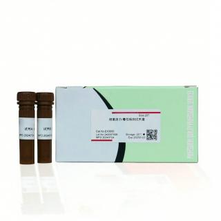
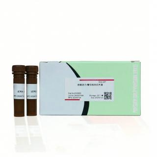
Cell Stimulation KIt(With Protein Transport Inhibitor) KCEA-1
Name: Cell Stimulation KIt(With Protein Transport Inhibitor)
Specification; 4*100ul
Storage conditions: Store at -20℃,6 months
Shipping conditions: Freeze transportation
Product Introduction
In cells, activation of PKC can cause the phosphorylation of many downstream protein kinases, forming a cascade reaction, leading to the expression of a variety of Proteins, which in turn causes cell activation. PKC can be activated by the combined action of DAG and Ca2+. PMA is a potent PKC activator; Ionomycin is a potent, selective calcium ionophore. PMA is often used in combination with Ionomycin to stimulate immune cell activation and is used to induce the production of cytokines in in vitro cell cultures.
At the same time, the protein transport inhibitor Monensin ( is a commonly used protein transport inhibitor that can destroy the Golgi apparatus and inhibit intracellular protein transport. It is often used to inhibit the secretion of secreted Proteins such as cytokines so that they remain within the cell. ) or Brefeldin A ( a specific inhibitor of protein transport that can block the transport of secreted Proteins and membrane Proteins from the endoplasmic reticulum to the Golgi apparatus.) to inhibit the exocytosis of cytokines to accurately analyze the expression levels of cytokines. If immunodetection of secreted Proteins is performed (eg, ELISA, Western blot, or multiplex protein assay), it is not necessary to treat the cells with Monensin or Brefeldin A.
Treatment with PMA and ionomycin is sufficient to induce activation and cytokine production in many cell types. BrefeldinA and Monensin cause accumulation of secreted proteins in the endoplasmic reticulum and Golgi apparatus. Therefore, this Kit can be used for the induction of cytokines and other secreted proteins in in vitro cultured and isolated cells and subsequent intracellular detection. For unstimulated samples, Brefeldin A and Monensin (CAT: IK-PTI-1) can be directly selected as controls.

Experimental case ( for reference only )
In Vitro
Cell
Flow Cytometric Analysis of the Percentages of Th17 Cells.(Spleen Mononuclear Cell/SMC, Thyroid Mononuclear Cell/TMC)
Phorbol 12-myristate 13-acetate (PMA, Solarbio) and Ca-ionomycin (Solarbio) were added into the 12-well plates containing the SMC or TMC suspension, and brefeldin A (BFA, Solarbio) was added after 1 h followed by incubation for 3 h (total stimulation of 4 h). Cells were then collected, washed, and surface stained with APC-labeled CD4 antibody at 4℃ in the dark for 30 min. IC fixation buffer was then added followed by incubation at 4℃ in the dark for 20 min. Finally, cells were washed, resuspended in permeabilization buffer, and stained with PE-labeled IL-17A antibody at 4℃ in the dark for 30 min. Flow cytometric analyses were performed using a FACScanto flow cytometer.
 sales
sales